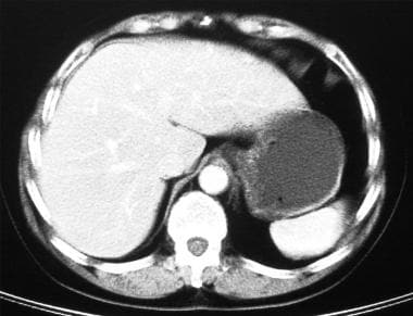
Exceptions to this rule include radiopaque foreign bodies and gross bowel obstruction, as well as moderate to large amounts of pneumoperitoneum. Although x-ray can distinguish among these according to principles discussed in more detail in Chapter 5, most pathologic processes are not well differentiated from normal findings by x-ray. g., bone, vascular calcifications, and ureteral calcifications). The abdomen contains four principal densities: air, fat, water (soft tissues and fluids such as blood), and calcified structures (e. Unfortunately, x-ray has limited utility in abdominal imaging. These are elaborated upon in later sections on individual forms of abdominal pathology.Ībdominal x-ray was the first abdominal imaging modality, used shortly after Roentgen’s discovery of x-ray in 1895. We review each of these modalities here, pointing out some general principles about their diagnostic capabilities and limitations. Fluoroscopy for gastrointestinal (GI) studies and abdominal angiography have seen a decline in use with the increasing availability and diagnostic utility of CT scan for the same applications. Nuclear scintigraphy and MRI are more rarely employed for selected conditions. The most commonly used emergency department abdominal imaging modalities include plain x-ray, CT, and ultrasound. Mesenteric angiography for gastrointestinal bleeding Gastrointestinal bleeding, tagged red cell study Intestinal foreign body, macroparasite, CT Pancreatitis and complications including pseudocyst, CT Inflammatory bowel disease and colitis, CTĬholelithiasis, cholecystitis, and gallbladder polyps, ultrasoundīiliary tract obstruction and choledocholithiasis, CT Tissue densities on CT and fat as a contrast agentĩ-33 see also sigmoid and cecal volvulus CT For quick reference, Table 9-1 lists the content of figures in this chapter. In some cases, multiple modalities from the same patient are displayed together, because they depict a clinical story that can reveal strengths and weaknesses of various imaging modalities. We refer to these chapters rather than repeating their content, recognizing that the clinical presentations of abdominal conditions can be quite nonspecific and that abdominal imaging sometimes must be selected to include an array of possible pathology, including traumatic, vascular, and genitourinary abnormalities.įigures in this chapter are organized in general by type of pathology. Chapter 16 delves into interventional radiologic procedures, including many for abdominal conditions.

We conclude with a comprehensive survey of important abdominal pathology, describing the diagnostic accuracy and imaging findings for each of the commonly employed modalities for each condition.Ĭhapter 10, Chapter 11, Chapter 12 review the imaging of abdominal trauma, abdominal vascular conditions, and genitourinary conditions, respectively. We describe a systematic approach to interpretation of abdominal x-ray and CT and a focused approach to abdominal ultrasound interpretation. In this chapter, we discuss the strengths, limitations, and indications for common emergency abdominal imaging modalities. Ideally, imaging should also reliably identify patients who do not require any urgent or emergent therapy and can safely be managed as outpatients.

Abdominal imaging must identify immediate life-threats such as leaking abdominal aortic aneurysm, conditions requiring urgent surgical intervention such as appendicitis, and conditions requiring antibiotic therapy such as diverticulitis without abscess. By 2005, 8% of adult patients visiting one tertiary care emergency department underwent abdominal computed tomography (CT). According to the National Hospital Ambulatory Medical Care Survey, about 8% of emergency department visits are for complaints potentially relating to the abdomen, including vomiting and abdominal pain. Abdominal imaging is essential to clinical decision making in emergency medicine.


 0 kommentar(er)
